Bone tissue is a highly dynamic and diverse tissue, both structurally and functionally. It has an ability to perform a wide array of functions, and responds to a variety of metabolic, physical, and endocrine stimuli. For majority of injuries, bone has the self-healing capability for forming scar-free tissue. However, there are injuries that may result with delayed union or non-union, where bone regeneration is required. At present, several surgical methods exist to treat bone defects, including Ilizarov intercalary bone transport
method, Masquelete technique, growth factor stimulation, and bone replacement. Application of these methods has so far resulted in relatively successful outcomes. Unfortunately, these methods come with noteworthy disadvantages including lengthy treatment, adverse effects on patient’s psychology, repeated surgical procedures, and often long hospitalization times. Collectively, these drawbacks and limitations associated with surgical techniques make bone substitutes (grafts or scaffolds) an attractive alternative.
At BIREL, we fabricate (3D print) layered polymer structures for bone defects, characterize these structure to compare with native bone as well as test their biocompatibility.


Tissue harvesting. Sectioned distal femur (A, B), the whole femur (C), and proximal femur (D, E). Specimen dimension: 2 cm × 2 cm × 2 cm (A, E).
3D-printed scaffolds with different pore sizes.

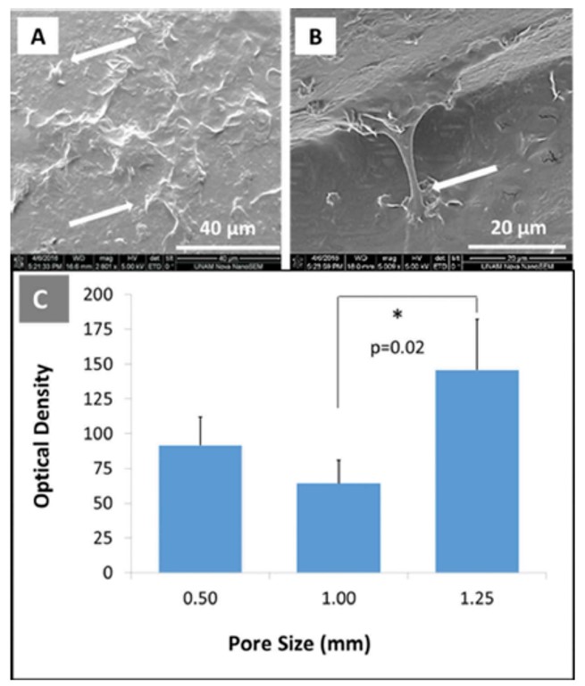
SEM micrographs obtained from 3D-printed scaffolds (pore diameter = 1.00 mm), demonstrating (A, B) attachment of hBMSC at day 3, and (C) cell proliferation at day 7. Arrows point to the adhered cells. * indicates significant difference at p < 0.05.
Source: Velioglu et al., Connective Tissue Research 2019, 60 (3): 274–282.

